Intervertebral Disc Disease


|
When Good Discs Go Bad
By Dr. Becker Intervertebral disc disease is a serious condition seen more often in dogs than cats. Intervertebral discs are cushioning pads of fibrocartilage that sit between most of the vertebra of the spinal column. The discs have an outer layer of tough fibrous tissue and a center that is more of a gel-like substance. They act as shock absorbers for the bones called vertebra in the spinal column. Unfortunately, intervertebral discs are subject to degeneration, bulging outward, and even bursting or rupturing. When something goes wrong with a disc, the material inside escapes into the spinal column, pressing against the spinal cord or nerve roots, which causes pain, nerve damage, and sometimes, paralysis. This is the condition known as intervertebral disc disease or IVDD. Depending on the location of the damaged disc, problems can occur anywhere in the animal’s body from the neck to the rear limbs. In cats, the problem discs are more often found in the neck and upper back. In humans, the condition is sometimes called a slipped disc or a herniated disc. IVDD is one of the most common neurologic disorders seen in pets, especially dogs. Most aging dogs have some degeneration of intervertebral discs, which commonly results in a condition known as spondylosis. |

Two Types of Intervertebral Disc Disease
There are two forms of IVDD in dogs -- Hansen Type I and Hansen Type II. Hansen Type I is the acute, explosive herniation of a disc. This type of IVDD is typically seen in middle-aged chondrodystrophic dogs with breed-specific inherited skeletal deformities lurking in their DNA. These dogs include the Dachshund, Shih tzu, Beagle, Pekingese, Poodle, Corgi, Basset Hound, and others with characteristics of genetic dwarfism. Hansen Type II IVDD involves a gradual progressive protrusion of disc material that affects non-chondrodystrophic breeds who are older, usually between the ages of 8 and 10. This form of IVDD is most commonly seen in German Shepherds, Labrador Retrievers, and Dobermans. Obese and out-of-shape dogs of predisposed breeds are at much higher risk of acquiring IVDD. The average age of kitties who develop the condition is 8 to 10 years. |
|
Symptoms of IVDD
There are a number of symptoms that can occur with intervertebral disc disease, but they’re also seen with other conditions, which is why an accurate diagnosis is needed. Signs you should watch for include reluctance to move the neck and head or a lowered head -- some animals only move their eyes to look at you. They don’t want to move their head or neck because it’s painful. An animal holding his head low and shifting just his eyes to look at you is definitely suspicious for IVDD. Other symptoms can include back pain, stiffness, crying out unexpectedly when touched or while moving, abdominal tenderness or tenseness, an arched back or hunched posture, incomplete or inappropriate urination, dragging one or more legs, toeing or knuckling over when walking or standing, weakness, stiffness, an odd or tentative gait, reluctance to sit or stand, or an unwillingness to jump. Some dogs have anxiety, because they know it’s going to hurt when they move. Other signs: reduced appetite or activity level, loss of bowel or bladder control, trembling or shaking, a loss of general coordination, paralysis in one or more limbs, or, in a worst-case scenario, sudden collapse. |

Diagnosing IVDD
A complete neurologic exam should be performed to help identify the location of the injured disc. Regular X-rays may show a problem area in the spine, but the spinal cord and the actual discs don’t appear on X-rays, so special imaging may be required. X-rays can be helpful in ruling out other potential causes of spinal cord damage, including tumors occurring in the bone, fractures, discospondylitis, and discospondylosis. A myelogram, which requires general anesthesia, is a procedure in which a dye is injected into the spine, making it visible when X-rayed. A herniated disc or spinal cord compression can usually be visualized with this particular technique. A spinal tap to assess cerebral spinal fluid is sometimes used to rule out other causes of symptoms such as infection, inflammation from autoimmune disease, or cancer. The best method for visualizing the spinal cord – and if you’re planning to do surgery, certainly a necessary step – would be an MRI. An MRI is a non-invasive technique that produces the most useful images of the spine, spinal cord, nerve roots, and intervertebral discs. The earlier IVDD is diagnosed and treated, the better your pet’s chances of avoiding a permanently painful and sometimes paralyzing condition. |

|
Treatment Options
Once a diagnosis of IVDD has been made and the affected discs located, a treatment plan can be developed. The goals for treating IVDD patients are to eliminate pressure on the spinal cord and resolve inflammation in order to return the pet to a pain-free and fully mobile life. Treatment can involve medical management or surgical intervention, depending on the severity of the disease. If the dog no longer has mobility and has lost deep pain sensation, the connection from the brain to the body has been severely compromised. Unfortunately, the only way to attempt to re-establish the connection is with surgery. Time is of the essence in these situations, and delaying surgery even for 24 hours can often dramatically reduce the possibility of a positive outcome. If surgery is necessary, there are a variety of techniques that can be used. The goal of any procedure is to relieve pressure on the spinal cord at the site of the damaged disc. In acute cases where the animal still has some mobility and a superficial pain response -- which indicates there’s still a viable connection between the brain and the body -- pain management and control of inflammation should be accomplished first. In any case, once a pet’s pain and neurological symptoms are well-controlled either medically or surgically, there will need to be an extensive period of complete rest in order for healing to occur. All disc patients must have a well-padded bedding area -- a small area they can’t get up and move around in. If the animal can’t reposition herself on her own, it’s important to turn her every few hours to prevent bedsores. Assistance with urination, defecation, eating, and drinking is often also necessary. 
|
|
Importance of Physical Therapy for IVDD Patients
During this time of complete rest, there are some very important therapies that can speed healing and improve your pet’s chances of a successful outcome. Acupuncture and electroacupuncture, which is sending a microcurrent of electricity to and from acupuncture points (which are really big nerve bundles), can be very beneficial at helping to re-establish the nerve connections in the body. Massage with or without medical-grade therapeutic essential oils is very good for disc patients as well. Massage of limbs and axial muscles not directly involved with the site of the injury and passive range of motion exercises can help improve circulation and assist with lymphatic drainage. Physical rehab technicians are trained to use gentle joint compressions to help maintain patient comfort and reduce pain. Also, these techniques help to maintain limb strength and muscle mass. Laser therapy at the surgical site or over the area of injury will promote a more rapid healing response, and neuromuscular electrostimulation will help slow muscle atrophy from disuse. When healing is far enough along, underwater treadmill therapy or swim therapy is an absolutely amazing tool for helping the body recover from neurologic trauma. As patients continue to improve, a land treadmill enhances endurance and improves gait and movement patterning. Physio balls and specific therapeutic exercises can be utilized to improve limb strength and core stability. Cavaletti poles are very effective for improving proprioceptive input and coordination. All that to say, pets who undergo rehabilitation after treatment for intervertebral disc disease heal faster, with a far better long term outcome than pets treated with medical or surgical intervention alone. The good news is most pets who maintain deep pain sensation through an episode of this disease can be well managed without surgery, especially in cases where the condition is diagnosed and treated early. Unfortunately, IVDD symptoms recur in about 50 percent of pets, especially if they are obese, out of shape, or if they’re allowed to jump freely. This is why regular physical therapy that focuses on establishing and maintaining core strength and muscle tone reduces the risk of recurrence, and helps keep disc patients’ quality of life excellent. |
|
Intervertebral discs are located between the vertebrae (bones of the spine). Each disc has two parts, a fibrous outer layer and the jelly-like interior. When disc herniation occurs, the interior either protrudes (bulges) or extrudes (ruptures) into the vertebral canal, where the spinal cord resides. The onset of herniations can be either acute or chronic. When the spinal cord is compressed by this disc material, the dog or cat experiences signs ranging from mild back or neck pain to paralysis, loss of sensation, and loss of bladder and bowel control. Disc disease can be serious; in some cases, paralysis or fecal and urinary incontinence may be permanent.
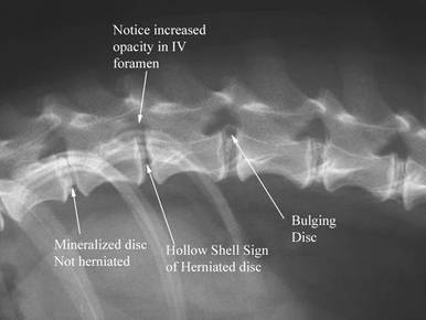
When discs mineralize, the mineralization starts centrally. When they become completely mineralized, they look a lot like a lens end on (like the one on the left in the x-ray image). If a mineralized disc herniates, it will leave a hollow shell or a partially hollow shell. Because mineralization starts centrally, a hollow shell is a sign of herniation. Note that this disc seen above has an increased opacity (looks whiter) in the IV foramen. Intervertebral disk herniations are most common in the long, low chondrodystrophic breeds (e.g., Dachshund, Basset Hound, Beagle, Cocker Spaniel, Shih Tzu, Lhasa Apso, Pekingese, and Corgi). It is a genetic predisposition due to the animal’s conformation. These low-slung dogs tend to get the bulging extrusions. Large breed dogs are more typically affected with protrusions. The degeneration weakens the disk, allowing it to herniate. However, disk herniations can also be caused by physical trauma (an accident, such as being hit by a car), or the onset of a disease (such as cancer). Intervertebral disk disease sometimes occurs in cats, but it is not as common as it is in dogs. Discs in the neck can have the same types of problems as do the disks in the back. If the herniations are mild to moderate, they cause only neck pain or a forelimb limp; if severe, all four limbs may be paralyzed. In affected dogs of chondrodystrophic (long, low-slung) breeds, disk degeneration occurs within the first few months of life, but the actual herniation typically occurs suddenly at around 3 to 6 years of age. In non-chondrodystrophic breeds, the disk degeneration usually starts at age five, with the herniation occurring slowly over time (6 to 8 years of age). |

Grading of Clinical Signs and Diagnosis
A neurological examination allows the severity of clinical signs to be graded as follows: Grade 5: normal Grade 4: ambulatory, but mildly paraparetic (weak/wobbly) Grade 3: markedly paraparetic (weak/wobbly), but is able to get up on his/her own Grade 2: severely paraparetic (weak/wobbly); good voluntary motion still present in hindlimbs, but cannot get up without assistance Grade 1: slight voluntary limb motion present Grade 0: paraplegic (no voluntary motion present). This grade is further subdivided as to whether or not deep pain sensation is present. Diagnosis may require radiographs of the spine, myelography (a type of imaging involving the injection of a contrast agent [a liquid that x-rays don’t go through] into the spinal canal to pinpoint the compressed area of spinal cord), CT or MRI scan, or other tests. A spinal tap under general anesthesia may be required to examine the cerebrospinal fluid for signs of other diseases. Treatment and Prognosis Mild cases may be managed medically. Confinement to a crate with minimal physical activity (no jumping, no running, no going up/down stairs, no playing, etc.) is necessary for several weeks. Pain medication, such as steroid-like drugs, muscle relaxants, or anti-inflammatories may be prescribed on a short-term basis. Paralyzed or chronically affected dogs usually require surgery, but the success of the surgery will vary, depending on the amount of damage that the spinal cord has incurred. More than 90% of the dogs who have the ability to sense pain in their hindlimbs will walk again after surgery; only about 60% of the dogs who have lost deep pain sensation will walk again. This surgery may require the expertise of a veterinary surgical specialist or neurosurgeon. Pet owners need to provide intensive care for the pet, no matter which treatment is used. Full recovery usually takes weeks to months. The prognosis depends on the clinical signs, how long the problem has been present, and how the dog responds to treatment. Some animals respond to the medical/surgical treatment, and some end up with permanent paralysis and fecal/urinary incontinence. The majority of dogs treated appropriately do well long term. Roger L. Welton, DVM Founder and Chief Editor, Web-DVM.net President, Maybeck Animal Hospital Article updated 8/19/2012 http://www.web-dvm.net/intervertebraldiscdisease.html |


|
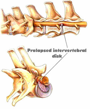
Description
Intervertebral discs are flexible structures between individual vertebrae. Disc prolapse describes the pathological changes of intervertebral discs and their overall negative effect on the spinal chord. Affected dogs often show difficulties leaping onto higher ground, i.e., the couch, or any act that places strain on the vertebral column. Climbing stairs or jumping in general may also be impaired. Being handled along the spine or lifted from the ground is often painful. A dropped head is a common sign of dogs with disc problems. In very severe or advanced cases the dog’s hindquarter can become paralyzed. As a result, affected dogs collapse onto their hind legs and lose control of their anal sphincter, which causes an involuntary bowel movement to occur. Paralysis of the front legs may also develop if the prolapsed disc is located in the neck. As a result of the degenerative processes the intervertebral discs lose flexibility. As a consequence of losing flexibility, they may rupture as a result of physical strain or trauma. The fragments from ruptured discs then apply pressure to the spinal cord, and this may damage nervous tissue. Spinal segments under high mechanical strain are more prone to prolapse. Causes of the condition are often breed-related. Dachshunds, Pekinese, Scottish Terriers, Springer Spaniels or Poodles are commonly affected. Even in early years they may develop prolapsed disc. Otherwise, the condition is often a result of the aging process and is often seen in older animals. Some pathological processes of the spinal cord may be detected by an x-ray. However, definite diagnosis can only be obtained from an MRI. Conservative treatment with anti-inflammatory drugs is curative in mild cases. However, a severe disc prolapse requires surgery and the removal of problematic disc fragments. If you suspect that your dog has a prolapsed disc, consult a veterinary clinic. Dogs suffering from a prolapsed disc should be lifted carefully into cars, onto couches, or over stairs. For a harness or restraint, chest gear should be used in favor of a collar. Excessive weight is putting extra pressure on the vertebral column and should be reduced as much as possible. http://www.petsicon.de/dog/diseases/prolapsed-disc |


Group on Facebook
|

|